Functional Magnetic Resonance Imaging Reveals Taiji’s Real-time Effects on Neuronal Networks of the Brain
Keywords:
Tai Chi, fMRIAbstract
Background
Taiji has beneficial effects on physical and mental health. While various traditional metrics have been applied to assess physical improvement from taiji, measuring changes in the brain to ascertain the etiology of benefits to mental health is not as straightforward. Recently, functional magnetic resonance imaging (fMRI) has been applied to the study of taiji. In this study, we measure real-time changes of neuronal networks reflecting mental taiji exercise inside an MRI scanner.
Methods
On four different days, fMRI data were acquired from a healthy volunteer with expertise in taiji. During each session, four sets of 4D fMRI data were obtained alternating between the pure resting and mentally practicing taiji states. Data were analyzed to identify intrinsic connectivity networks and brain regions that demonstrated significant difference in functional connectivity between pure-resting and mental taiji-practicing conditions.
Results
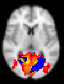
Differences in functional connectivity between pure-resting and mental taiji-practicing conditions were found in the visual, sensorimotor and default mode networks. Specifically, in regard to the default mode network (DMN), the precuneus and right angular gyrus showed relatively decreased activity during the mental taiji-practicing condition.
Conclusions
fMRI signal changes in the visual, sensorimotor and default mode networks during the mental practice of taiji may have implications for uncovering the neural mechanisms underlying the benefits of taiji.
Published
How to Cite
Issue
Section
Copyright (c) 2020 Phillip Kuo, Xintuo Zhang, Carol Stuehm, Ying-hui Chou, Nan-kuei Chen

This work is licensed under a Creative Commons Attribution-NonCommercial-NoDerivatives 4.0 International License.
